This web page was produced as an assignment for Genetics 677, an undergraduate course at UW-Madison
Scientific Article Review
Mueller’s Cadherins and mechanotransduction by hair cells very thoroughly outlines cadherins 23 and 15 in many contexts of the mechanotransduction pathway. The article begins with a factually rich description of the mechanosensory hair cells (see figure 1). The article then discusses mutative cadherin proteins and their link to Usher’s syndrome. Mueller hints at the greater extracellular domains of the two (23 and 15) as reasons for their importance in transduction. Cadherin 23 and 15 are part of two non-classical cadherin families, FAT and protocadherin, respectively. Figure two serves this section by contributing a visual comparison against a classical cadherin. The appearance of other deft-causing diseases is also brought up.
The next two sections discuss CDH23 and PCDH15 in normal developing and matured hair cells. This section delves into much detail about the different isoforms found distinctly in stereocilia, but the take-home message is that novel forms of both proteins can be seen solely in the stereocilia, which suggests some post transcriptional/translational evolutionary adaptation. A large discussion ensues as the author plays devil’s advocate, questioning whether we can actually say these are linkage components, the main point being that we are ignorant of the stereocilia linkage type (see figure 3).
Figures 4 and 5 encompass the next section of the piece, which presents the newest relevant data that the protein harmonin forms a binding complex with CDH23 and PCDH15, and these complexes are what interact with other signal pathway components, thereby necessitating harmonin as a component of mechanotransduction.
The last section discusses another protein group, myosin motor proteins, as a possible active transporter of cadherins to the cytoplasmic membrane where they are integrated into. Studies have shown that mice with a novel myosin mutation Myo7a go deaf, and observation of these hair cells show structure similar to those with defective cadherin and harmonin proteins.
The article as a whole is rigorously informative, and provides a good scope of research of , and relating to, cadherins in stereocilia.
Article Figures
Figure 1: Hair cell bundling in stereocilia
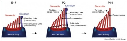
Shows 3 diagrams representing the development of a hair bundle on the tip of a hair cell body, found in the cochlea. Sterocilia are arranged smallest to largest with a kinocilian at the top (the 'final straw'). As the bundle grows the height proportions are maintained and tip links composed of CDH23 and PCDH15 proteins are produced. These links, along with other intersterocilia links, serve to efficiently transfer sound impulses to the kinocilia, to transduce the signal.
Figure 3: two models for beginnning mechanotransduction
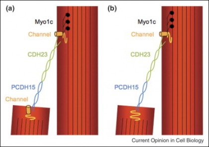
Two working models of molecular tip link models in sterocili. Both indicated CDH23 and PCDH15 proteins working in line with one another to transfer physical stimuli. Model A suggests that mechanotransduction pathways occur in both sterocili (channel heads are found on both), whereas Model B suggests the pathway to be conserved to only one of the sterocili.
Figure 4: downstream interactions from CDH23
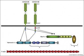
Notice the different functions of CDH23 in signal transduction. This is due to a difference in structure, so within different cadherins of the mechanotransduction pathway there are isoforms of each.
Figure 5: Downstream signaling of the PCDH15-CD1 complex
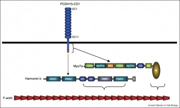
Harmonin interacts with the PCDH15-CD1 complex (this is not a fully accepted depiction) and Myo7a binds to the complex.
Reference:
Ulrich Müller. Cadherins and mechanotransduction by hair cells Current Opinion in Cell Biology
Volume 20 Issue 5, October 2008, pages 557-566
Volume 20 Issue 5, October 2008, pages 557-566
Ben Hofeld, [email protected], last updated: 5.15.2010, Link to course page:www.gen677.weebly.com
